LYMPHATIC RECONSTRUCTION FOR BRAIN DISEASE
What evidence supports existence of glymphatic and meningeal lymphatic pathways?
Clinical studies and publications are summarized herein by MMI and are not intended to mislead or omit information.
Meningeal lymphatic drainage: novel insights into central nervous system disease
Focus: This paper is a review article focusing on the significant role of meningeal lymphatic drainage in central nervous system (CNS) diseases.
Method: This paper is a review article.
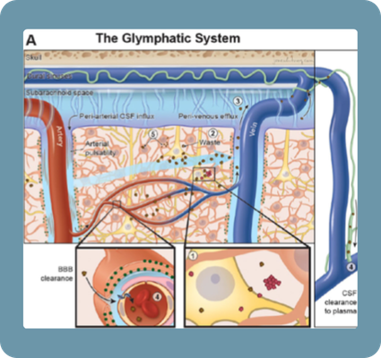
Finding: Meningeal lymphatic vessels play a crucial role in draining CSF, metabolic byproducts (such as amyloid-β proteins), and immune cells from the CNS, and their dysfunction is increasingly recognized as a significant factor in the pathogenesis and progression of various CNS diseases, including neurodegenerative disorders, stroke, and traumatic brain injury.
Limitation: Understanding of regulatory and damage mechanisms of meningeal lymphatics under physiological and pathological conditions is currently limited.
CSF transport at the brain–meningeal border: effects on neurological health and disease
Focus: New evidence regarding CSF transport mechanisms at the brain–meningeal border, including glymphatic and meningeal lymphatic systems, and their implications for neurological health and diseases.
Method: Review article synthesizing current knowledge on CSF transport.
Finding: “The existence of specialized structures that allow a continuous exchange of CSF between different anatomical compartments at the brain–meningeal border is challenging conventional notions around molecular transport within the brain.“
Limitation: The precise mechanisms of CSF-to-dura mater transport and immune cell trafficking remain poorly understood, particularly in humans.
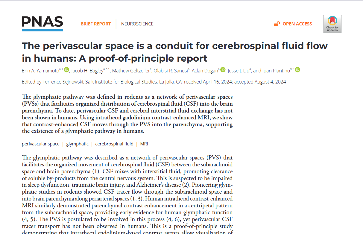
The perivascular space is a conduit for cerebrospinal fluid flow in humans: A proof-of-principle report
Focus: Provide proof-of-principle evidence that the perivascular space in humans functions as a conduit for CSF flow into the brain parenchyma.
Method: Contrast agent administers intrathecally in 5 neurosurgical patients. Two postcontrast brain MRIs performed at 12, 24, or 48 hours.
Finding: CSF flows through perivascular spaces into the brain parenchyma, confirming the existence of a glymphatic pathway in humans.
Limitation: Small sample size.
MRI imaging provides evidence of glymphatic drainage from human brain to cervical lymph nodes

Focus: CNS lymphatic drainage to cervical lymph nodes in humans in-vivo.
Method: MRI following intrathecal contrast agent administration.
Finding: “The time course of lymph node enhancement coincided with brain glymphatic enhancement rather than with CSF enhancement.”
Limitation: Measurement errors may arise from image noise and partial averaging effects, despite efforts to minimize them.
The glymphatic system clears amyloid beta and tau from brain to plasma in humans
Focus: Investigating whether sleep-active glymphatic function contributes to the overnight clearance of amyloid beta (Aβ) and tau from the human brain.
Method: A clinical randomized crossover study where participants underwent both overnight normal sleep and overnight sleep deprivation conditions. Plasma AD biomarkers measured at evening and morning timepoints.
Finding: Sleep-active glymphatic function significantly enhances the overnight clearance of Aβ and tau from the human brain into plasma in healthy individuals.
Limitation: The study included a large number of regressors given the relatively small number of amyloid-positive participants.
Navigating Unchartered (Neuroimmune) Waters

Dr. John Kipnis
Reveals how meningeal lymphatics, cerebrospinal fluid (CSF) dynamics, immune signaling, and sleep-dependent clearance mechanisms are integral to brain health, aging, and neurodegenerative diseases.
Cerebrospinal fluid-mediated brain clearance: insights from human studies
Focus: This study focuses on cerebrospinal fluid (CSF)-mediated brain clearance, particularly insights gained from human studies. It investigates the essential role of brain fluids, primarily CSF and interstitial fluid exchange, in removing waste products from brain metabolism and by-products of brain injury.
Method: Intrathecal Contrast-Enhanced Magnetic Resonance Imaging (gMRI), Phase-Contrast MRI, CSF Clearance Test (Blood-based).
Finding: CSF is essential for brain clearance, exchanging with interstitial fluid to remove waste products. Impaired clearance leads to the accumulation and aggregation of proteins, a hallmark of neurodegenerative and dementia diseases.
Limitation: A key question remains unanswered: to what extent does CSF-mediated brain clearance contribute to overall brain solute clearance compared to local proteolytic degradation and active transport over the blood-brain barrier in various brain regions.
Brain-wide glymphatic enhancement and clearance in humans assessed with MRI
Focus: The study’s primary focus is to investigate the extent to which the subarachnoid cerebrospinal fluid (CSF) compartment directly communicates with the extravascular compartment of human brain tissue.
Method: This study was a prospective and observational investigation that examined the distribution and clearance of a CSF tracer in the human brain using MRI.
Finding: The study provides in vivo evidence, for the first time, of brain-wide CSF tracer enhancement and clearance in humans at a rate suggesting a significant role for bulk flow and vascular pulsations rather than pure diffusion. It also showed a delayed tracer clearance in patients with dementia.
Limitation: Assessment of cerebral clearance with MRI by absolute quantities is not possible at the current level, as the normalized T1 signal cannot be assumed to be strictly proportional to the amount of contrast agent in each image voxel.
Acute sleep loss decreases CSF-to-blood clearance of Alzheimer’s disease biomarkers
Focus: The study’s focus is to determine the effect of acute sleep loss on Alzheimer’s disease (AD) biomarkers in plasma, specifically amyloid beta (Aβ) and tau.
Method: This study utilized a cross-over design involving five cognitively normal, amyloid-negative participants. Each participant underwent both a sleep-deprived condition and a normal sleep control condition, separated by 4-6 months. During both conditions, cerebrospinal fluid (CSF) and blood samples were collected every 2 hours over a 36-hour period using venous and lumbar catheters.
Finding: A key finding of the study is that one night of sleep deprivation has opposing effects on AD biomarkers in CSF and plasma. This suggests that sleep loss decreases the clearance of these biomarkers from the central nervous system and disrupts their transport between CSF and blood.
Limitation: A small number of participants. All five participants were cognitively normal and amyloid-negative, which prevents the extension of these results to individuals with amyloid deposition, cognitive impairment, or medical comorbidities that might affect brain clearance or alter plasma AD biomarkers.
Glymphatic dysfunction in Alzheimer’s disease: A critical appraisal
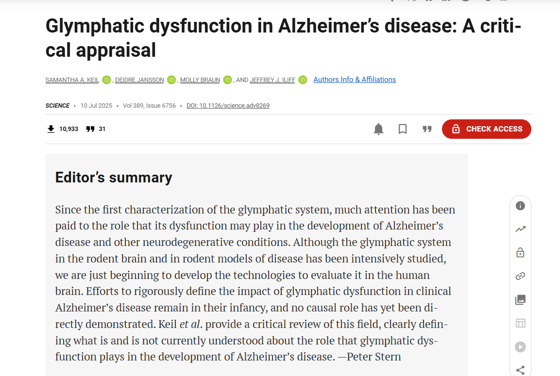
Focus: The study critically appraises the role of glymphatic dysfunction in Alzheimer’s disease (AD), differentiating between findings from animal studies and human clinical research.
Method: This study is a critical appraisal and review article that synthesizes and evaluates existing scientific literature on the role of glymphatic dysfunction in AD.
Finding: Despite strong evidence from preclinical rodent studies suggesting that glymphatic impairment contributes to the development of AD pathology, a clear causal role for glymphatic impairment in the development of clinical AD in human populations has not yet been established.
Limitation: A significant limitation highlighted in the study is the difficulty of directly but noninvasively measuring glymphatic function in the human brain.
Explore questions and research
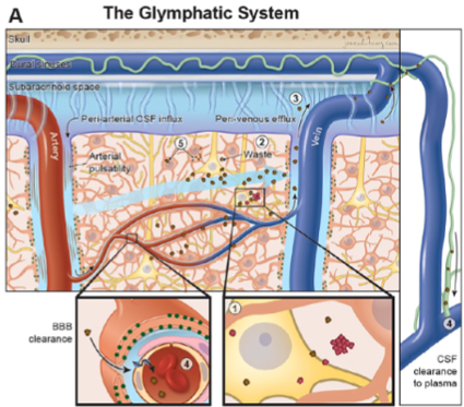
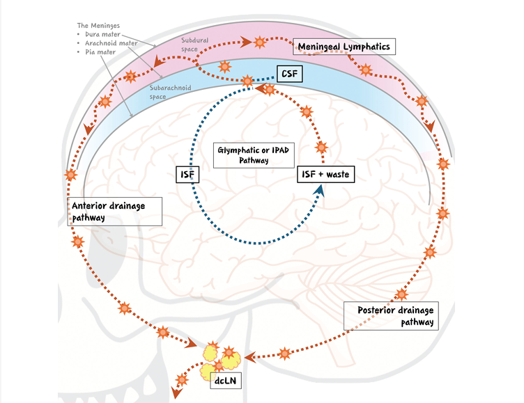
What evidence supports existence of glymphatic and meningeal lymphatic pathways?
Image Source: Nature
Is there a mechanical problem to be addressed?
Image Source: JNM
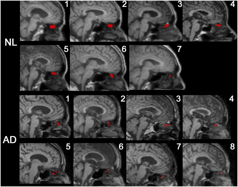
Is there a causal relationship between protein levels and cognitive/functional decline in Alzheimer’s disease patients?
Image Source: NIH News
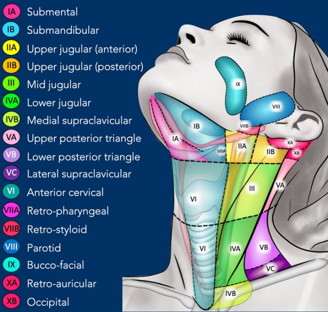
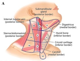
What is the anatomic target for a mechanical solution?
Image Source: The Radiology Assistant: Cervical Lymph Node Map
What’s the proposed procedure and should it be done in a supermicro fashion?
Image Source: APS
Is there evidence demonstrating the impact of the LVA/LNVA procedure in the neck?
Image Source: International Journal of Surgery
Pharmacologic treatments and severity distribution for Alzheimer’s disease
Image Source: NIH News
The information provided is solely for educational purposes relating to scientific information on the brain lymphatic system. Such summaries are not intended to mislead or omit information. Clinical studies and publications may be summarized herein. Click on each resource to access the full publication.
The safety and effectiveness of the Symani Surgical System for lymphatic reconstruction for brain disease has NOT been established by any regulatory agency.
The Symani Surgical System is indicated in the U.S. for soft tissue manipulation in open microsurgical procedures to perform:
- Anastomosis, suturing, and ligation microsurgery techniques on small blood vessels and lymphatic ducts between 0.1 and 2.5mm in open free-flap surgery of the breast, mouth, scalp, and extremities and in open lymphatic surgery of the extremities.
- Dissection of soft tissues.
Clinical indications vary by geography. Refer to global indications for more information. For risks, cautions, warnings, and full prescribing information, please reference Symani safety and performance information.
