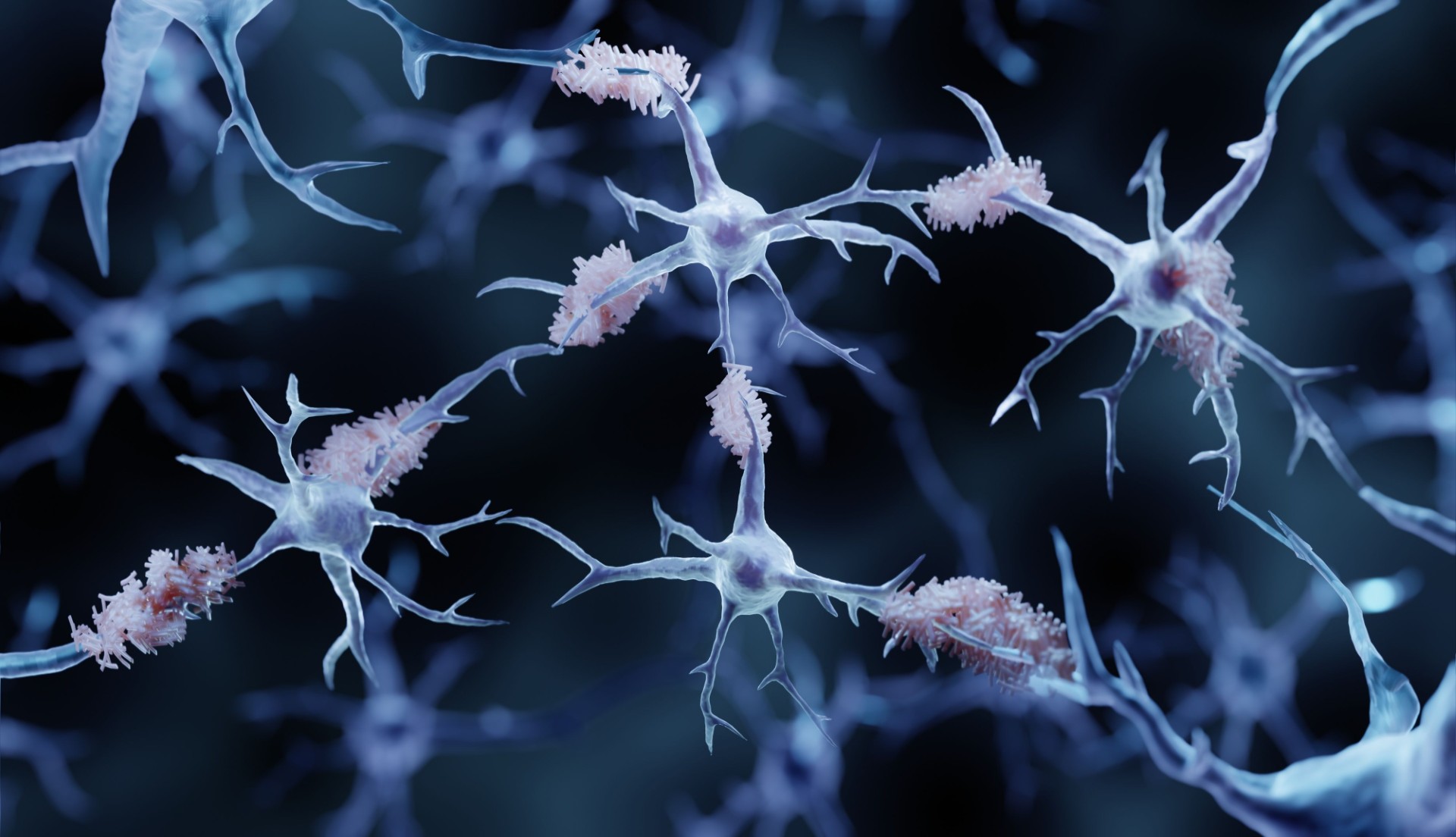
LYMPHATIC RECONSTRUCTION FOR BRAIN DISEASE
Is there a mechanical problem to be addressed?
Clinical studies and publications are summarized herein by MMI and are not intended to mislead or omit information.
Age and amyloid effects on human central nervous system amyloid-beta kinetics
Focus: The study focused on determining the relationship between age, amyloidosis, and amyloid-beta (Aβ) kinetics in the human central nervous system (CNS).
Method: The study utilized stable isotope labeling kinetics (SILK) to analyze Aβ turnover. Participants received a 9-hour intravenous infusion of leucine to label newly synthesized proteins, including Aβ isoforms. Plasma and CSF samples were collected for 36 hours and the incorporation of labeled leucine into soluble Aβ was quantified. A compartmental model was applied to determine kinetic parameters of Aβ turnover.
Finding: A key finding of the study is that the fractional turnover rate of soluble Aβ peptides in the CNS slows significantly with age.
Limitation: Independent comparisons regarding ApoE allele genotype effects on Aβ kinetics could not be made because ApoE4 was highly associated with amyloidosis, preventing researchers from isolating the specific impact of ApoE on Aβ metabolism before amyloidosis occurred.
Cerebrospinal fluid clearance in Alzheimer’s disease measured with dynamic PET
Focus: Relationship between CSF clearance, Aβ accumulation and cognitive function in individuals with AD.
Method: Two PET tracers to estimate CSF clearance.
Finding: “Ventricular CSF clearance measures were correlated with reductions in Alzheimer’s disease (AD) of 18 and 27% compared to normal.”
Limitation: The absence of direct, non-invasive methods limits understanding of Aβ clearance in humans.
Decreased CSF clearance and increased brain amyloid in Alzheimer’s disease
Focus: Relationship between CSF clearance, Aβ accumulation and cognitive function in individuals with AD.
Method: Two PET tracers to estimate CSF clearance.
Finding: “Ventricular CSF clearance measures were correlated with reductions in AD of 18 and 27% compared to normal.”
Limitation: The absence of direct, non-invasive methods limits understanding of Aβ clearance in humans.
Brain glymphatic fluid mapping in Alzheimer’s disease: a human MRI and PET study
Focus: This study focuses on establishing parenchymal CSF fraction (pCSF) mapping as a sensitive and clinically feasible biomarker for evaluating glymphatic fluid distribution in AD. Ultimately, the research seeks to advance the monitoring of AD and predict responses to treatment by identifying a reliable biomarker for glymphatic clearance.
Method: Retrospective study involved 29 subjects (16 cognitively normal, 13 MCI/AD) who underwent both MRI and C-PiB PET scans. Three MRI-based measures of glymphatic structural alterations were compared. C-PiB PET was used to measure Aβ deposition. The study evaluated cross-correlations among these MRI biomarkers and their respective associations with amyloid deposition via linear regression, adjusting for age and sex.
Finding: Parenchymal CSF (pCSF) mapping demonstrates a significantly stronger positive association with Aβ deposition compared to diffusion-based free-water mapping and perivascular space burden, even after adjusting for age and sex.
Limitation: One study limitation is its cross-sectional nature, which restricts the ability to draw causal inferences between glymphatic dysfunction and the progression of AD. Additionally, the sample size, while adequate for preliminary findings, may not fully capture the nuanced effects of glymphatic clearance across various stages of the disease.
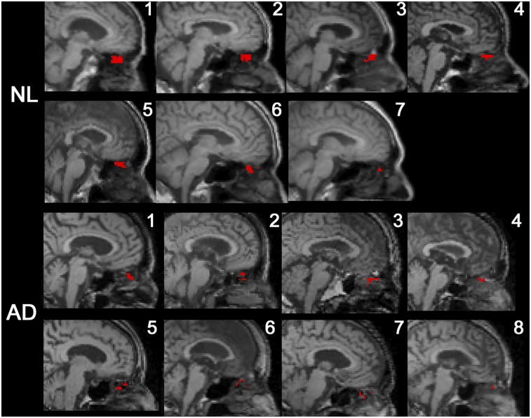
Structural and vascular alterations of deep cervical lymph nodes in amyloid PET-positive Alzheimer’s disease patients
Focus: Assessing the structural and vascular alterations of deep cervical lymph nodes (dCLNs) in patients with amyloid PET-positive AD using high-frequency color Doppler ultrasound (HFCDU).
Method: Cross-sectional design involving 25 amyloid PET-positive AD patients and 25 age-matched cognitively normal controls. Participants underwent HFCDU of dCLNs in neck zones 2–5.
Finding: “HFCDU demonstrated distinct morphological and vascular abnormalities of dCLNs in AD, particularly in Zone 5, suggesting impaired lymphatic drainage contributes to AD pathology and supporting lymphatic imaging as a potential biomarker for the failure of lymphatic drainage of the brain.”
Limitation: The study was conducted on a relatively small number of patients at limited stages of AD, which may restrict generalizability.

Explore questions and research
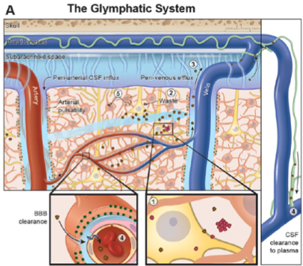
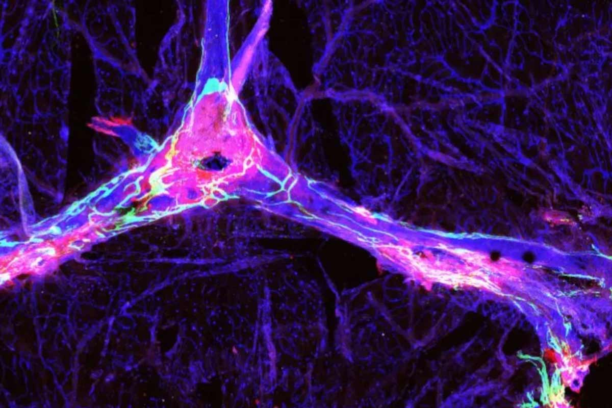
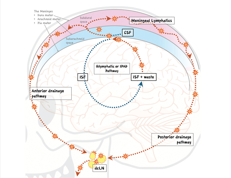
What evidence supports existence of glymphatic and meningeal lymphatic pathways?
Image Source: Nature
Is there a mechanical problem to be addressed?
Image Source: JNM
Is there a causal relationship between protein levels and cognitive/functional decline in Alzheimer’s disease patients?
Image Source: NIH News
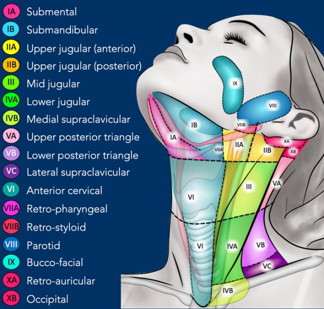
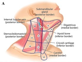
What is the anatomic target for a mechanical solution?
Image Source: The Radiology Assistant: Cervical Lymph Node Map
What’s the proposed procedure and should it be done in a supermicro fashion?
Image Source: APS
Is there evidence demonstrating the impact of the LVA/LNVA procedure in the neck?
Image Source: International Journal of Surgery
Pharmacologic treatments and severity distribution for Alzheimer’s disease
Image Source: NIH News
The information provided is solely for educational purposes relating to scientific information on the brain lymphatic system. Such summaries are not intended to mislead or omit information. Clinical studies and publications may be summarized herein. Click on each resource to access the full publication.
The safety and effectiveness of the Symani® Surgical System for lymphatic reconstruction for brain disease has NOT been established by any regulatory agency.
The Symani Surgical System is indicated in the U.S. for soft tissue manipulation in open microsurgical procedures to perform:
- Anastomosis, suturing, and ligation microsurgery techniques on small blood vessels and lymphatic ducts between 0.1 and 2.5mm in open free-flap surgery of the breast, mouth, scalp, and extremities and in open lymphatic surgery of the extremities.
- Dissection of soft tissues.
Clinical indications vary by geography. Refer to global indications for more information. For risks, cautions, warnings, and full prescribing information, please reference Symani safety and performance information.
